
An echocardiogram, a heart ultrasound, is a non-invasive test that uses sound waves to create images of the heart. Doctors order this common test to evaluate the structure and function of the heart and detect any abnormalities or diseases.
Why is an echocardiogram done?
A heart ultrasound helps in diagnosing a variety of heart conditions, including:
1. Heart defects: An echocardiogram detects structural abnormalities, including holes in the heart or narrowed heart valves.
2. Heart failure: This test helps determine the extent of damage to the heart muscle and how well the heart pumps blood.
3. Cardiomyopathy: A heart ultrasound detects abnormalities in the thickness or shape of the heart muscle.
4. Heart valve disease: This test helps detect abnormalities in the heart valves, such as stenosis or regurgitation.
5. Pericardial disease: An echocardiogram can detect any inflammation or fluid buildup around the heart.
How is an echocardiogram done?
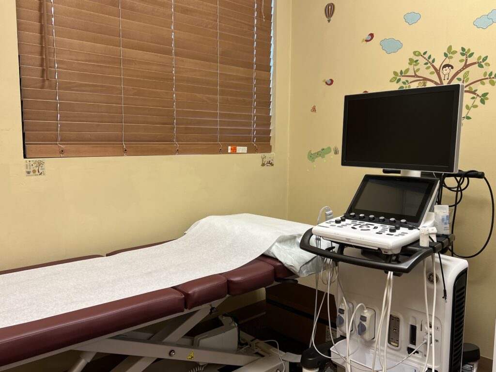
An echocardiogram is a painless and non-invasive test that typically takes 45-60 minutes to complete. Here’s what you can expect during the test:
1. You will be asked to remove your clothing from the waist up and lie on your back on an examination table.
2. A technician will apply a gel to your chest and then place a transducer on different areas of your chest, which emits sound waves.
3. The transducer will send sound waves through your chest, bouncing back and creating images of your heart on a monitor.
4. The technician will move the transducer around your chest to get different heart views.
5. After the test, the technician will wipe off the gel from your chest.
Types of echocardiograms
There are several types of echocardiograms, including:
1. Transthoracic echocardiogram (TTE): This is the most common type of echocardiogram, which involves placing the transducer on the chest.
2. Transesophageal echocardiogram (TEE): This test involves placing a thin tube with a transducer at the end in your mouth and down your throat to get closer images of your heart.
3. Stress echocardiogram: This test is done to evaluate how well your heart functions under stress, such as during exercise.
4. 3D echocardiogram: This test provides a detailed 3D image of your heart, which can help detect any abnormalities.
What are the Risks of a Heart Ultrasound?
An echocardiogram is a safe and non-invasive test, and there are typically no associated risks. However, in rare cases, some people may experience an allergic reaction to the gel used during the test.
In conclusion, it is a simple and painless test that can provide valuable information about the heart’s structure and function. If you’re experiencing any symptoms of heart disease or have any risk factors, talk to your doctor about whether an echocardiogram is right for you.
Here at Peds Happy Hearts, Dr. Hua performs and reads the echocardiogram. You get the result on the same day. To schedule the heart ultrasound, contact our office at 406-272-2376.
3 thoughts on “Echocardiogram: Understanding the Heart Test”